Left Hip Muscles Anatomy / Anatomy Of Knee. This mri hip joint axial cross sectional anatomy tool is absolutely free to use. The six hip adductor muscles are all located in the adductor or medial compartment of the thigh and all mainly adduct the thigh at the hip joint. Injury to the iliopsoas may cause hip pain and limited mobility. Hip pain explained will teach you about the anatomy of the hips and pelvic area and how many different types of body tissues interact. The strong muscles of the hip region also help to hold the hip joint together and prevent dislocation.
Some of the other muscles in the hip are: Hip muscle anatomy is a complex topic. The bones of the hip include the femur, the ilium, the ischium, and the pubis. Related posts of muscles of the lower back and hip diagram neck muscle anatomy ultrasound. The muscles of the neck can be divided into groups according to their location.
Hip Flexor Strain Symptoms Causes And Treatment from post.medicalnewstoday.com The hip muscles include pelvic and groin muscles. Left hip muscles anatomy : Hip extension and internal rotation of left hip joint in the final phase of the gait cycle. The different bursae of the hip region (trochanteric, ischial and iliopectineal bursae) Learn about hip muscles human anatomy with free interactive flashcards. Muscle construction with label 12 photos of the muscle construction with label , human muscles. One at the left hip, and one at the right hip. They are important for stabilising the body and for moving the legs.
These are gracilis, pectineus, adductor longus, adductor brevis, adductor magnus, and adductor minimus muscles.
One at the left hip, and one at the right hip. The strong muscles of the hip region also help to hold the hip joint together and prevent dislocation. A bursa that sometimes causes problems in the hip is sandwiched between the bump on the outer hip (the greater trochanter) and the muscles and tendons that cross over the bump. They are important for stabilising the body and for moving the legs. These muscles include the gluteus maximus muscle (the largest muscle in the body) and the hamstrings group, which consists of the biceps femoris, semimembranosus, and semitendinosus muscles. These are gracilis, pectineus, adductor longus, adductor brevis, adductor magnus, and adductor minimus muscles. The muscles of the neck can be divided into groups according to. The hip's essential muscles are the sartorius, rectus femoris, gluteus minimus and medius, iliopsoas, adductors, and hamstrings. The hamstrings are three muscles at the back of the thigh that affect hip and knee. The hip muscles are composed of multiple flexors, extensors, adductors, abductors, and rotators that work together. The femur is a long bone that consists of a head, neck, shaft, and other protrusions such as the greater and lesser trochanter, which serve as attachment sites for muscles. Hip pain explained will teach you about the anatomy of the hips and pelvic area and how many different types of body tissues interact. The iliofemoral, pubofemoral, and ischiofemoral ligaments represent the thickenings of the joint capsule.
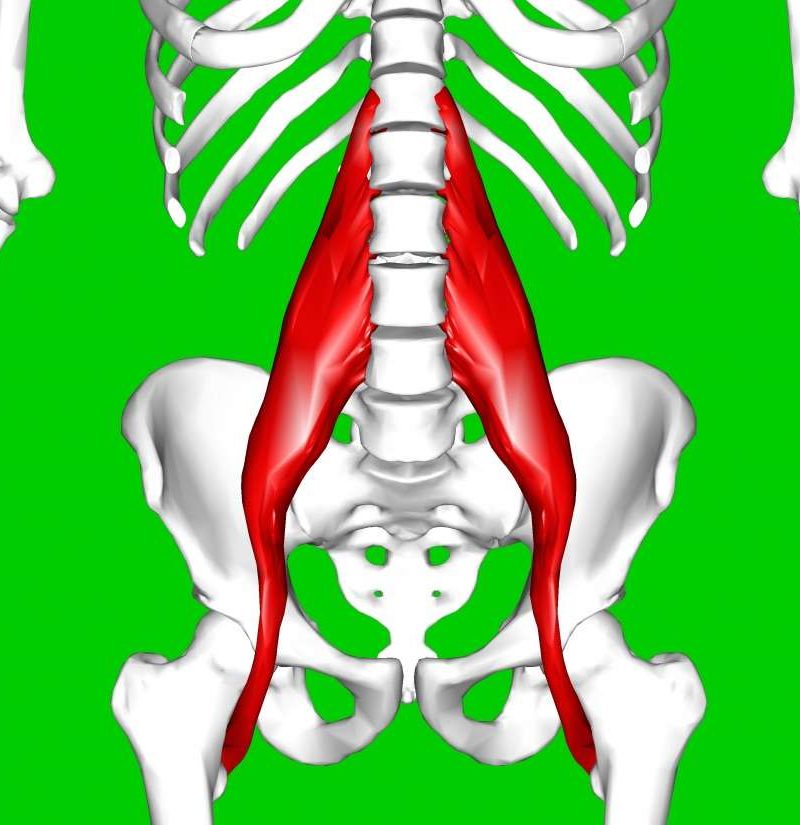
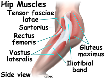
The view on the left has the rectus femoris cut away to show the vastus intermedius which is below it. This video also provides you with a. The hip joint is a ball and socket synovial joint, formed by an articulation between the pelvic acetabulum and the head of the femur. The neck muscles, including the sternocleidomastoid and the trapezius, are responsible for the gross motor movement in the muscular system of the head and neck. Iliopsoas muscle, a hip flexor muscle that attaches to the upper thigh bone.
Hip Anatomy Eorthopod Com from eorthopod.com Functionally, the hip joint enjoys a very high range of motion. The muscles of the neck can be divided into groups according to. Related posts of muscles of the lower back and hip diagram neck muscle anatomy ultrasound. Learn their anatomy efficiently and easily using kenhub's muscle anatomy and reference charts! Understanding the possible causes of hip flexor pain has a number of benefits. Here we explain the hip and groin muscles, their actions and exercises. The strong muscles of the hip region also help to hold the hip joint together and prevent dislocation. Rectus femoris muscle, one of.
Hip pain explained will teach you about the anatomy of the hips and pelvic area and how many different types of body tissues interact.
They are important for stabilising the body and for moving the legs. Rectus femoris muscle, one of. Muscle and tendon anatomy of the hip (adductors, gluteal muscles (or buttocks), hamstring muscles, femoral muscle quadrices). The pubis, ischium, and ilium together constitute the pelvis while the thigh bone is the femur. The hamstrings are three muscles at the back of the thigh that affect hip and knee. The muscles of the neck can be divided into groups according to their location. Related posts of muscles of the lower back and hip diagram neck muscle anatomy ultrasound. Left hip muscles anatomy : The hip itself is a ball and socket joint, much like the shoulder.the structures necessary to create this joint are the socket, the joint capsule, muscle, ligaments, and the neck. The thigh bone or femur and the pelvis join to form the hip joint. These muscles are the adductor longus, adductor brevis, adductor magnus, gracilis, and the obturator externus. The hip muscles include pelvic and groin muscles. The iliofemoral, pubofemoral, and ischiofemoral ligaments represent the thickenings of the joint capsule.
Related posts of muscles of the lower back and hip diagram neck muscle anatomy ultrasound. Rectus femoris muscle, one of. Attached to the bones of the skeletal system are about 700 named. Hip muscle anatomy is a complex topic. Left hip muscles anatomy / hip joint anatomy / related online courses on physioplus.
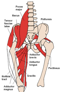
Muscles Of The Hip Wikipedia from upload.wikimedia.org Muscle and tendon anatomy of the hip (adductors, gluteal muscles (or buttocks), hamstring muscles, femoral muscle quadrices). Highly detailed 3d models, with textures up to 4k resolution, enable to examine the shape of each. Posterior view of gluteus maximus and gluteus medius in human anatomy, the muscles of the hip joint are those muscles that cause movement in the hip. The view on the left has the rectus femoris cut away to show the vastus intermedius which is below it. Muscle construction with label 12 photos of the muscle construction with label , human muscles. Anterior muscles extend your legs and flex your thighs. Iliopsoas muscle, a hip flexor muscle that attaches to the upper thigh bone. This video also provides you with a.
The bones together make up the hip.
Related posts of muscles of the lower back and hip diagram neck muscle anatomy ultrasound. The muscles that pull the legs together, such as those needed when riding a horse, are the adductor muscles of the hip.they originate at the pelvis and attach to the femur. Injury to the iliopsoas may cause hip pain and limited mobility. Attached to the bones of the skeletal system are about 700 named. Hip anatomy bones and joints of the hip. Left hip muscles anatomy : Rectus femoris muscle, one of. The anatomy of the fascia lata and iliotibial tract; Anatomy of the hip muscles. The hip's essential muscles are the sartorius, rectus femoris, gluteus minimus and medius, iliopsoas, adductors, and hamstrings. Highly detailed 3d models, with textures up to 4k resolution, enable to examine the shape of each. Blood vessels and nerves of the hip A bursa that sometimes causes problems in the hip is sandwiched between the bump on the outer hip (the greater trochanter) and the muscles and tendons that cross over the bump.
Share :
Post a Comment
for "Left Hip Muscles Anatomy / Anatomy Of Knee"

Post a Comment for "Left Hip Muscles Anatomy / Anatomy Of Knee"